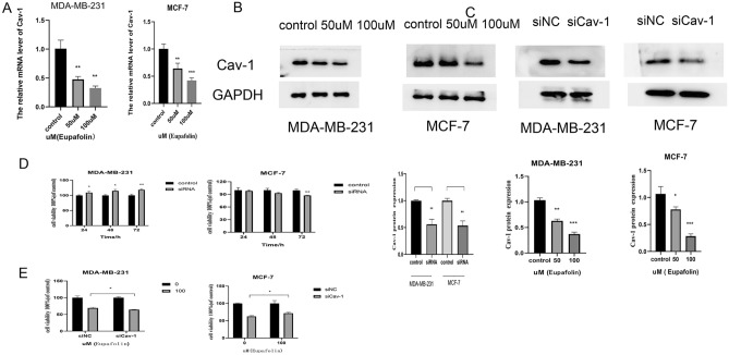
Figure 6.
Reduced Cav-1 is partially responsible for inhibition of proliferation of human breast cancer cells exposed to Eupafolin. (A,B) Total RNA and protein extracted from MDA-MB-231and MCF-7 cells treated with or without Eupafolin 50, and 100 μM for 24 h were used for quantitative real-time PCR and Western blot. Full-length images are presented in Supplementary Fig. 7. (C) MDA-MB-231and MCF-7 cells were transfected with Cav-1 siRNAs for 48 h and then subjected to Western Blot for evaluation of Cav-1 expression. Full-length images are presented in Supplementary Fig. 8. (D,E) MDA-MB-231and MCF-7 cells transfected with Cav-1 siRNAs or negative control siRNAs were treated with or without Eupafolin. Graphs of signal intensity were obtained through band densitometry and referred to GAPDH and control levels. Data are representative of three independent experiments and expressed as the mean ± SD. P > 0.05 indicates non-significance; ***P < 0.001; **P < 0.01; *P < 0.05.

