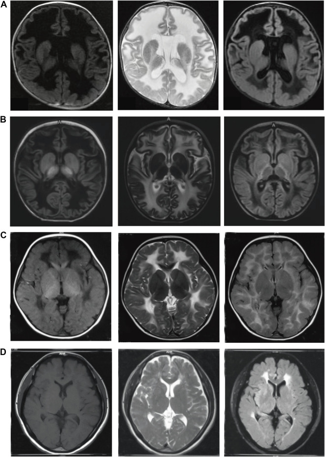
FIGURE 1.
Brain MRI of four patients with VWM at different onset ages. (A): case 1 with onset in neonatal period, the age at MRI was 3 months; (B): case 2 with onset in infancy, the age at onset was 11 months, the age at MRI was 18 months; (C): case 3 with onset in childhood, the age of onset was 3 years old, the age at MRI was 3 years old; (D): case 4 with onset in adulthood, the age of onset was 43 years old, the age at MRI was 43 years old. The left column:T1 weighted sequences (T1WI); The middle column:T2 weighted sequences (T2WI); The right column: T2 FLAIR sequences. The white matter showed low signal on T1WI, high signal on T2WI and low signal liquefaction on T2 FLAIR. The earlier the age of onset, the wider the range of white matter liquefaction, while the liquefaction sign was not evident in adult patients. Furthermore, the subcortical white matter was involved in the early stage.

