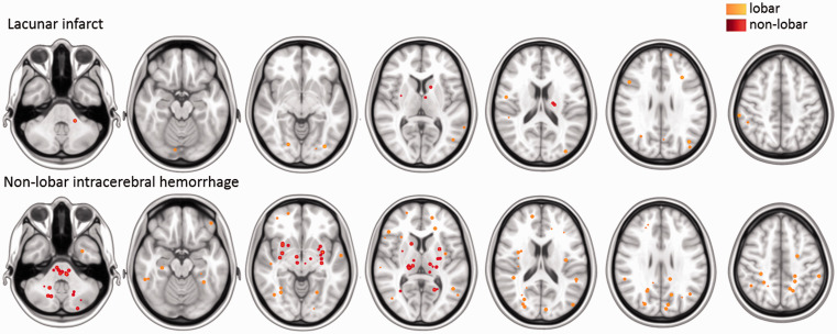
Figure 3.
Distribution of cerebral microbleeds in patients with lacunar stroke or non–lobar intracerebral hemorrhage. Spherical maps of cerebral microbleeds superimposed on a MNI–152 0.5 mm template with each sphere indicating a single microbleed, colour–coding represents lobar (orange) or non–lobar (red) locations.

