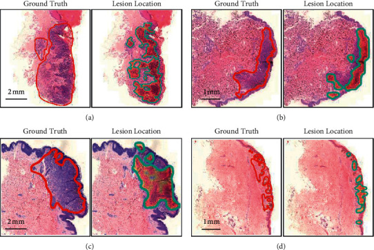
Figure 3.

Lesion location. The red line outlines the ground truth labelled by the pathologist and the heat map, with the blue line showing the lesion area located by the model. (a). Lesion location in melanoma WSI. (b). Lesion location in compound nevi WSI. (c). Lesion location in intradermal nevi WSI. (d). Lesion location in junctional nevi WSI.
