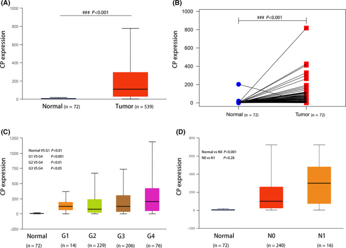
Fig. 6.

Overexpression of CP in ccRCC was correlated with lymph node metastasis stage and histological grade in the TCGA database. The mRNA expression level of CP in ccRCC tissues and normal renal tissues (A, B). Analysis of CP expression in ccRCC at different histological grades (C) and nodal metastasis status (D). Error bars represent standard deviation. P < 0.05 was as statistically significant.
