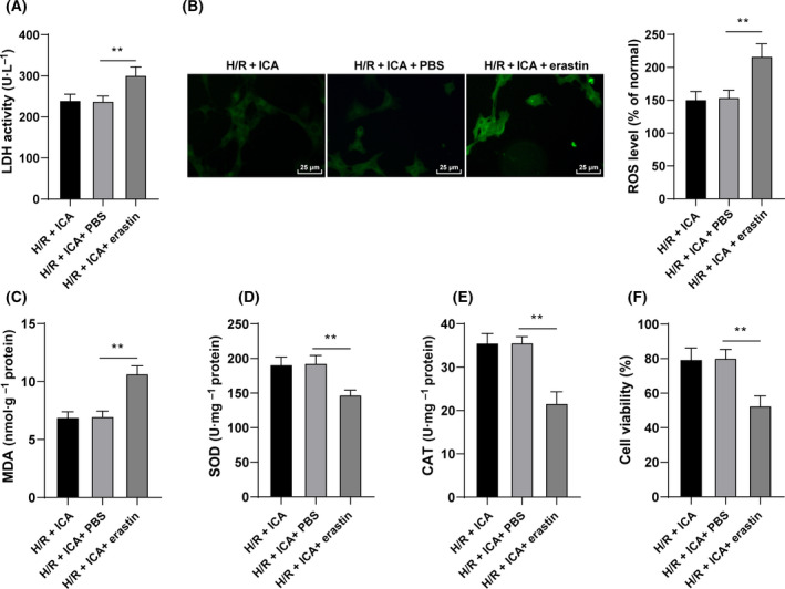
Fig. 3.

ICA exerted a protective effect on H/R‐induced cardiomyocytes by inhibiting ferroptosis. H/R‐induced cardiomyocytes were treated with 10 μm ICA and 5 μm erastin. (A) LDH content was detected. (B) Fluorescence intensity of ROS was detected using the DCFH‐DA probe. Scale bars, 25 μm. (C–E) The level of MDA and the activities of SOD and CAT were detected. (F) Cell viability was measured using CCK‐8 assay. The cell experiment was repeated three times. Data were presented as mean ± standard deviation. Data in (A)–(F) were analyzed using one‐way ANOVA, followed by Tukey's multiple comparison test, **P < 0.01.
