Figure 1.
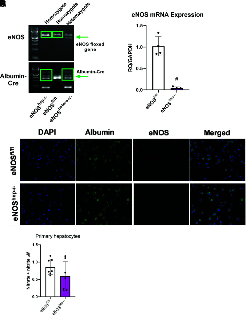
Confirmation of the deletion of hepatocellular eNOS in primary hepatocytes collected from both eNOSfl/fl and eNOShep−/− mice on a CD. A: Genotyping images displaying the floxed eNOS gene and the presence of the Albumin-Cre in our eNOSfl/fl and eNOShep−/− murine line. B: mRNA expression of eNOS from isolated primary hepatocytes (n = 4/group). C: Fluorescence microscopy of isolated primary hepatocytes, confirming the deletion of hepatocellular eNOS. Nuclei were stained with DAPI (blue), hepatocytes stained with albumin (green), and eNOS stained by anti-eNOS antibody (red) (n = 4–5/genotype). D: Nitrate and nitrite concentration in supernatant from isolated primary hepatocytes (P = 0.16; n = 6–8). Data are presented as mean ± SD. #Significantly different from eNOSfl/fl (P < 0.05). RQ, relative quotient.

