
Figure 6.
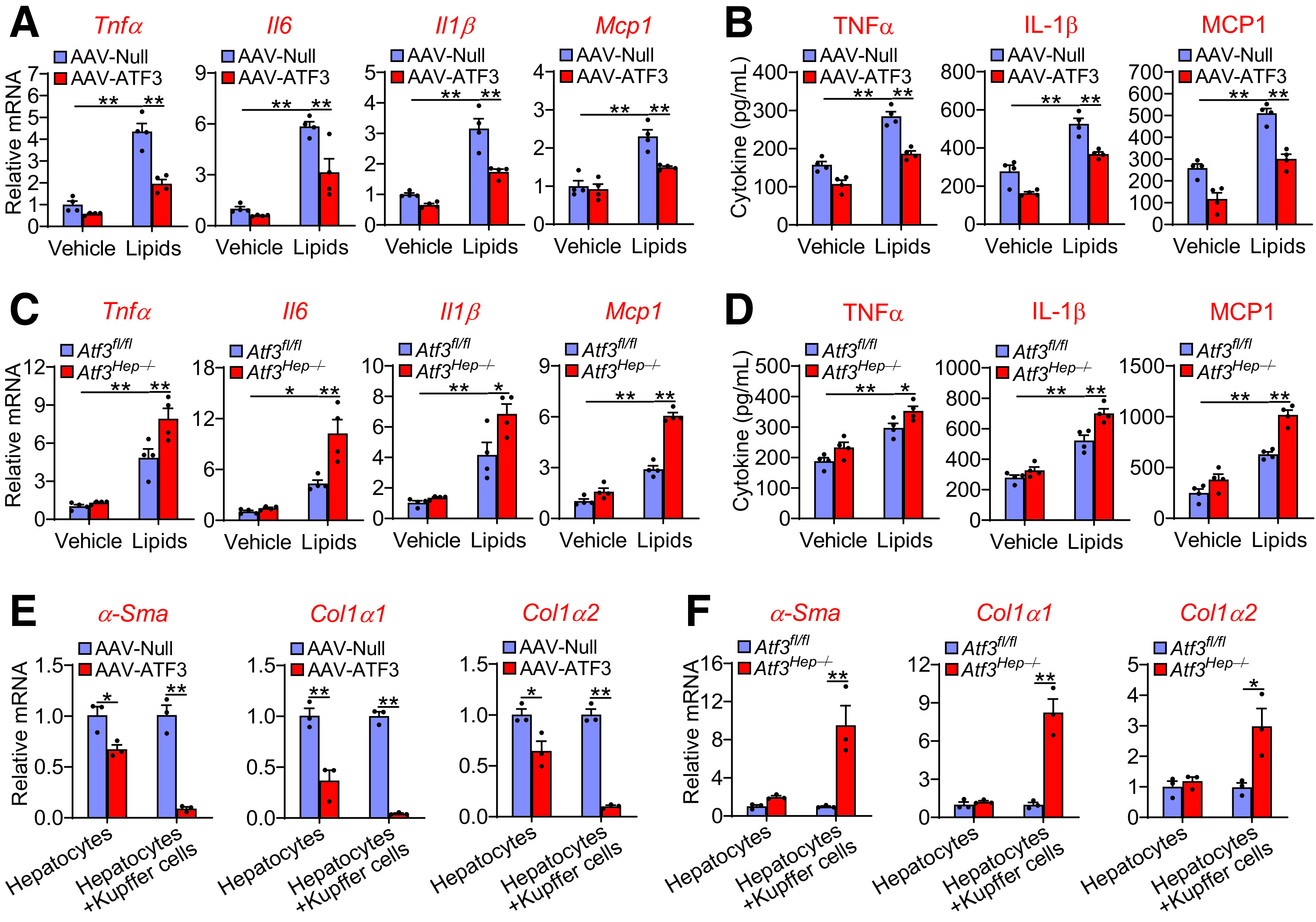
Hepatocytic ATF3 regulates Kupffer cell and stellate cell activation. A and B: Primary hepatocytes were isolated from mice injected i.v. with AAV8-ALB-Null or AAV8-ALB-ATF3 and then treated with vehicle or lipid mixture containing FFAs and cholesterol. After 24 h, the culture media were collected and used for treatment of Kupffer cells for 12 h. mRNA levels in Kupffer cells (A) as well as TNFα, IL-1β, and MCP1 levels in the media (B) were quantified (n = 4). C and D: Primary hepatocytes were isolated from Atf3fl/fl or Atf3Hep−/− mice and then treated with vehicle or lipid mixture for 24 h. The culture media were collected and used for treatment of Kupffer cells for 12 h. mRNA levels in Kupffer cells (C) and TNFα, IL-1β, and MCP1 levels in the media (D) were quantified (n = 4). E and F: Primary hepatocytes or primary hepatocyte plus Kupffer cells were incubated with lipid mixture for 24 h. The culture media were then used for treatment of stellate cells for 24 h, which were isolated from mice infected with AAV8-ALB-Null or AAV8-ALB-ATF3 (E) or Atf3fl/fl mice or Atf3Hep−/− mice (F). mRNA levels were determined (n = 3). All values are expressed as mean ± SEM. Two-way ANOVA was used for statistical analysis. *P < 0.05, **P < 0.01.