
Figure 1.
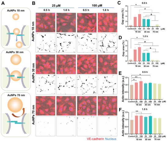
AuNPs‐induced endothelial leakiness and actin reorganization in HMVECs. A) Illustrated interactions between adherens junction and AuNPs of different sizes. VE–cadherin homophilic interactions among endothelial cells assist the connection and stability of the intact monolayer. The introduction of the different sized AuNPs affected the integrity of the adherens junction to different extents. The 18 and 30 nm AuNPs were small enough to migrate into the adherens junction and disrupt VE–cadherin homophilic interaction, while the 70 nm AuNPs were not able to cause significant disruption. B) Confocal fluorescence microscopy observed the occurrence of endothelial leakiness in the presence of different sizes (18, 30, and 70 nm) and concentrations (25 and 100 × 10−6 m) of AuNPs upon 0.5 and 1 h treatments. The images with black dots on a white background revealed the gaps’ distributions derived from the trainable Weka segmentation plugin in ImageJ software. Scale bar: 20 µm. C,D) Percentages of gaps area analyzed by ImageJ according to the gaps’ distribution images from panel B. E,F) Actin intensity analysis was performed by ImageJ software for the images in Figure S5 (Supporting Information). Data are shown as mean ± SD (n = 3), analyzed via two‐way ANOVA using GraphPad Prism 8, *, #, & represent P < 0.01 and ** represents P < 0.001 between the compared groups.