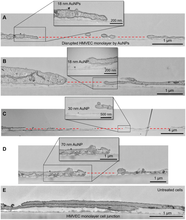
Figure 2.

Microtome transmission electron microscopy imaging of endothelial leakiness in HMVEC monolayers induced by AuNPs. A,B) Disruption of HMVEC monolayers by AuNPs of 18 nm in size after 30 min of exposure. Panel A shows the presence of an AuNP on the edge of cell membranes adjacent to the EL site. Panel B shows the presence of an AuNP inside the EL gap between two cells. C) EL between HMVECs induced by AuNPs of 30 nm in size. D) EL induced by AuNPs of 70 nm in size. E) Cell junctions between two HMVECs (control). The insets of panels A–D are zoomed in sections showing the presence of AuNPs. The EL sites are indicated by red dotted lines.
