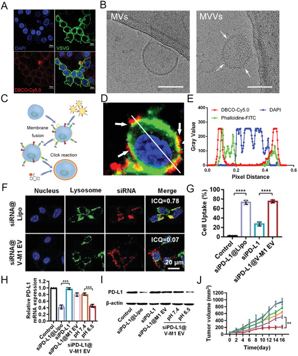
Figure 8.

Leveraging viral fusogen anchoring for membrane receptors’ transfer and siRNA delivery. A) Confocal images for VSVG overexpressing HEK293T cells after treatment with FITC‐VSVG‐tag antibody and DBCO‐Cy5.0. DAPI (blue); FITC (green); Cy5.0 (red). Scale bar: 10 µm. B) Cryo‐EM images of Bio‐MVs that from VSVG overexpressing HEK293T cells. White arrows show the virus‐like surface spike structures. Scale bar: 100 nm. C) Schematic illustration for the fusion procedure. D,E) Verification and characterization of membrane fusion behavior via confocal microscopy in vitro. DAPI = blue; DBCO‐Cy5.0 = red. Reproduced with permission.[ 120 ] Copyright 2020, Wiley‐VCH. F) Confocal observations of siRNA internalization mediated by Bio‐MVs that were genetically engineered with VSVG protein. Intensity correlation quotient (ICQ) analysis was performed to determine the colocalization. Scale bar: 20 µm. G–I) The delivering efficiency of siRNA tested with flow cytometry, RT‐qPCR, and western blot. J) Anticancer effects of siPD‐L1 encapsulating Bio‐MVs in vivo. Reproduced with permission.[ 121 ] Copyright 2020, Wiley‐VCH.
