
Figure 1.
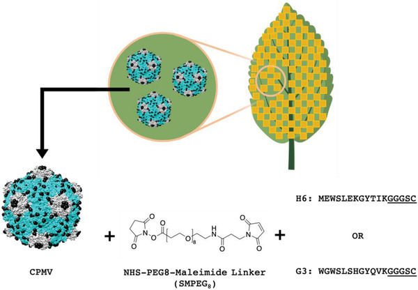
CPMV bioconjugation strategy. CPMV is first extracted from infected black‐eyed pea No. 5 plants. The large and small coat proteins are shown in blue and grey; surface exposed Lys side chains are highlighted as black spheres. The H6/G3 peptides with C‐terminal Cys side chain (the linker is underlined) were then conjugated to CPMV using an SMPEG8 linker via NHS‐maleimide chemistry. CPMV images and chemical structures were drawn with UCSF Chimera and ChemDraw software. The image of the leaf is created with BioRender.com.