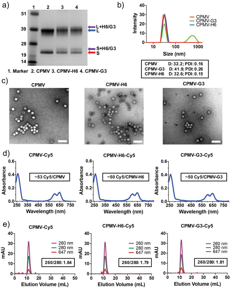
Figure 2.

Characterization of CPMV, peptide‐conjugated CPMV, and fluorescent CPMV particles. a) SDS‐PAGE of the CPMV particles. The purple arrows point to H6/G3 peptide‐modified coat proteins. The blue arrow points to the large coat protein (42 kDa), and the red arrow points to the small coat protein (24 kDa). b) DLS measurements of the CPMV particles. The box in black is displaying the average diameter in nm of the particles (D) and the polydispersity index (PDI). c) TEM images of uranyl acetate‐stained CPMV particles. Scale bars represent 100 nm. d) UV–vis of the fluorescent Cy5‐conjugated CPMV particles. The boxed insets are displaying the number of conjugated Cy5 particles per CPMV particle (based on Beer's Law). e) FPLC measurements of the fluorescent and peptide‐conjugated CPMV particles. The inset is indicating the 260/280 nm ratio at the peak of the FPLC curve. Corresponding CCMV data are shown in Figure S2e, Supporting Information.
