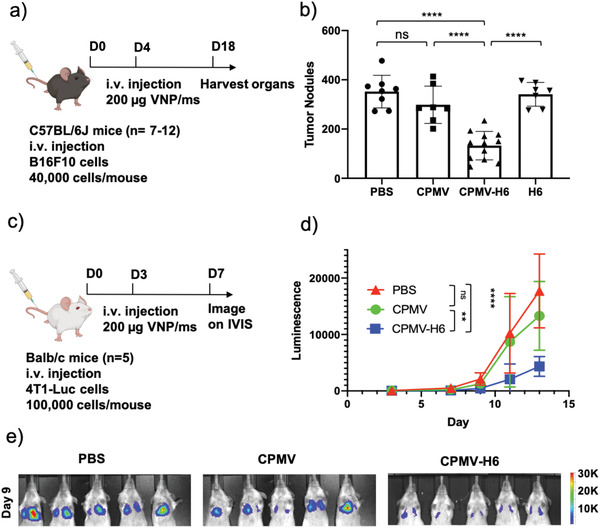
Figure 5.

S100A9‐targeted CPMV immunotherapy against lung metastasis from i.v. injected B16F10 melanoma and 4T1‐Luc breast cancer cells in mice. a) Treatment schedule of the metastatic B16F10 melanoma model using C57BL/6J mice and therapeutic administration of CPMV and CPMV‐H6. b) Quantitative analysis of tumor nodules counted in lungs harvested post‐treatment. c) Treatment schedule of the metastatic 4T1‐Luc breast cancer model using Balb/c mice. d) Quantitative luminescence of the tumors following region of interest (ROI) measurements of the images from (e). e) Luminescent imaging of the 4T1‐Luc tumors taken on the IVIS. The mice were imaged every two days following 150 mg kg−1 i.p. injection of D‐luciferin, and the luminescence was calculated using ROI measurements from the Living Image 3.0 software. One representative image taken on the IVIS on day 9 is shown. For B16F10 experiments an n = 7–12 animals per group and for the 4T1‐Luc experiments an n = 5 animals per group were assigned. Statistical significance was characterized as p < 0.05. All analyses were done by either one or two‐way ANOVA. * = p < 0.05, ** = p < 0.01, *** = p < 0.001, **** = p < 0.0001, ns = not significant. The image of the mouse is created with BioRender.com.
