
Figure 6.
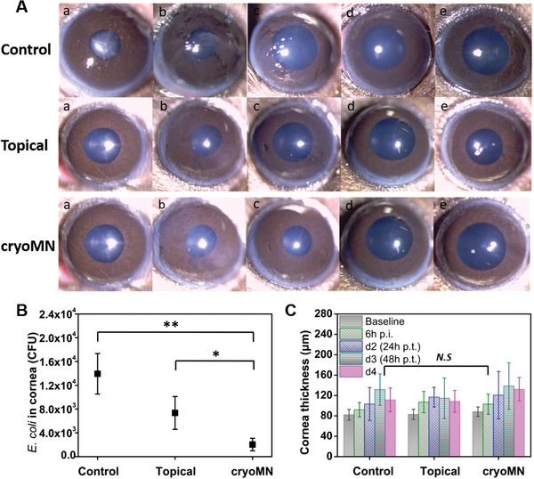
Ocular delivery of B. bacteriovorus with cryoMNs for eye infection: A) Cornea images taken by slit‐lamp photography (a. Baseline; b. 6 h p.i. after inoculation (prior treatment); c. Day 2 (24 h post treatment (p.t.)); d. Day 3; e. Day 4. B) Final E. coli concentration inside mouse corneas. N = 4. C) Cornea thickness before and after treatment every day. N = 4. * p < 0.1, ** p < 0.01, N.S means no significant difference.