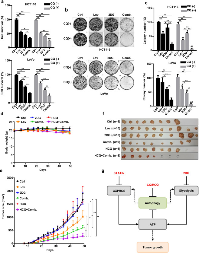
Fig. 6. The inhibition of autophagy increases the vulnerability of HCT116 cells and LoVo cells to combined treatment with lovastatin and 2DG.
a HCT116 and LoVo cells were treated with lovastatin (15 μM) and 2DG (5 mM) alone or in combination for 72 h in the presence or absence of chloroquine (CQ) (6.25 μM), and cell proliferation was determined (n = 3) (*P < 0.05, **P < 0.01. #P < 0.05, ##P < 0.01 versus indicated treatment group in the absence of CQ). b Colony formation of HCT116 and LoVo cells treated with lovastatin (5 μM) and 2DG (2 mM) alone or in combination in the presence or absence of CQ (400 nM) for 10 d. c Quantified data of (b) (n = 4). (*P < 0.05, **P < 0.01. #P < 0.05, ##P < 0.01 versus indicated treatment group in the absence of CQ). d HCT116 cells were injected subcutaneously into each hind limb of nude mice, and hydroxychloroquine (HCQ) (80 mg/kg every 2 d), lovastatin (25 mg/kg every 2 d), 2DG (5 mg/kg every 2 d), Comb. (lovastatin + 2DG) and HCQ + Comb. were administered orally. Ctrl n = 8, lovastatin n = 10, 2DG n = 10, HCQ n = 8, Comb. n = 9, HCQ + Comb. n = 8. Data were shown as the means ± SEM. e Tumor size in the mice shown in (d). *P < 0.05, **P < 0.01. Data are shown as the means ± SEM. f Tumors excised from mice in (d), and the dotted circle represents the tumor that completely disappeared after treatment. g Schematic representation of the combined effect of lovastatin and 2DG showing increased susceptibility of HCT116 cells to autophagy inhibition.

