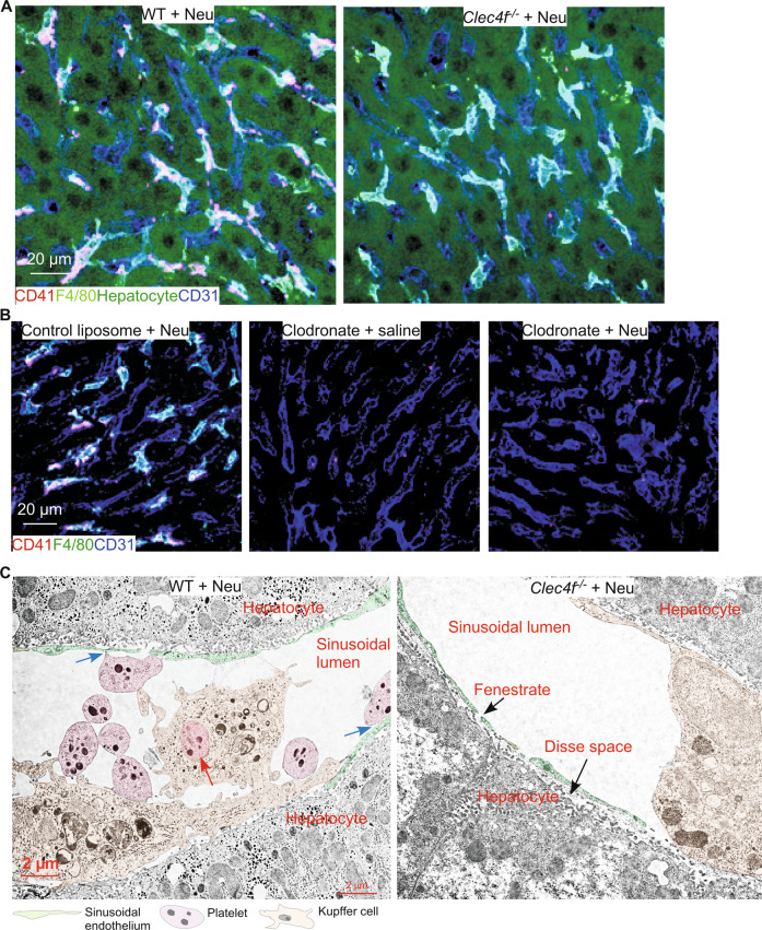
Fig. 4. Kupffer cells are the primary cell type phagocytizing desialylated platelets in the liver.
A, B Representative confocal microscopic images. CD41 marks platelets, F4/80 marks Kupffer cells, CD31 labels sinusoidal endothelium, and hepatocytes were detected by autofluorescence. C Pseudocolored TEM micrographs of WT or Clec4f−/− ultrathin sections. Red arrow marks a phagocytized platelet. Blue arrows mark potential interactions between platelets and sinusoidal endothelium.

