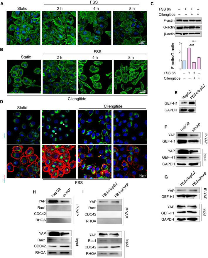
Fig. 6.

F‐actin transmits the biomechanical signal from integrin to YAP through GEF‐H1. (A) Cytoskeleton arrangement induced by FSS was examined using BODIPY (green stain) at the indicated time points after FSS treatment (n = 3). Scale bar: 10 μm. (B) Inhibition of the mechanotransduction of integrin induces the disruption of F‐actin (n = 3). Scale bar: 10 μm. (C) Western blotting analysis of the G‐actin/F‐actin ratio in HepG2, FSS‐HepG2, FSS+Cil‐HepG2 and Cil‐HepG2 cells (n = 3). Statistical analyses was performed by one‐way analysis of variance followed by Tukey test. Data are shown as mean ± SEM. ***P < 0.001. (D) Cilengitide induces the disruption of F‐actin (red) and reduces YAP (green) nuclear accumulation (n = 3). The arrows indicate the location of YAP. Scale bar: 10 μm. (E) Western blotting analysis of GEF‐H1 in HepG2 and FSS‐HepG2 cells. (F–I) Co‐IP analysis of control and YAP‐knockdown HepG2 cells subjected to FSS (n = 3).
