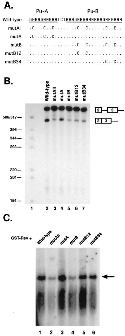
FIG. 5.
In vitro splicing and RNA binding of ESE mutants. (A) Sequence of two purine stretches (designated A and B) in exon 3. GAA repeats were mutated to GCA in the largest splicing construct (Fig. 2A) and RNA probe RREp4 (Fig. 4A). (B) In vitro splicing analysis of mutant ESE constructs. The locations of splicing products are indicated. Sizes are shown at the left (in nucleotides). (C) RNA gel mobility shift assays detecting GST-Rev binding to the mutant probes. The arrow points to the location of shifted RNAs.

