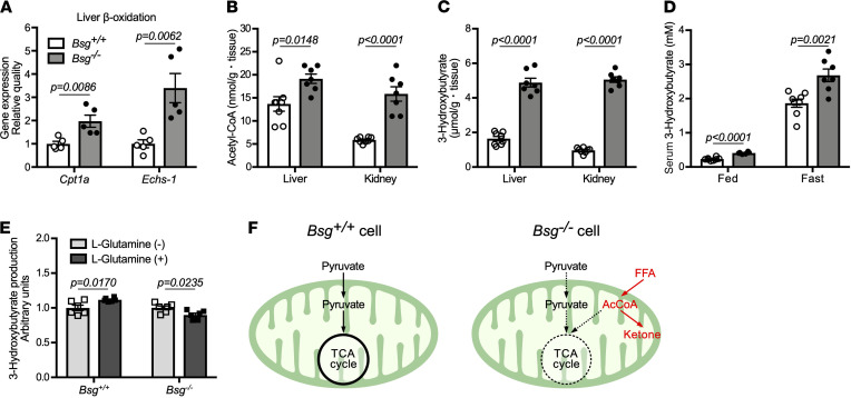
Figure 5. Increased ketogenesis via activation of fatty acid β-oxidation in Bsg deficiency.
(A) Concentrations of mRNAs encoding CPT1a and ECHS1 proteins, both of which are involved in fatty acid β-oxidation in the liver under fasting conditions (n = 5/genotype). mRNA levels were normalized to those encoding the housekeeping protein β-actin. White columns and circles, Bsg+/+ mice; gray columns and black circles, Bsg–/– mice. Scatter plots display the data for individual mice. (B and C) AcCoA (B) and the ketone body 3-hydroxybutyrate (C) contents in the livers and kidneys in fasting Bsg+/+ and Bsg–/– mice (n = 7–8/genotype). (D) Serum 3-hydroxybutyrate values in Bsg+/+ and Bsg–/– mice under feeding and fasting conditions (n = 6–8/genotype and condition combination). (E) 3-Hydroxybutyrate production by isolated hepatocytes derived from Bsg+/+ and Bsg–/– mice when the cells were cultured in the presence of l-glutamine (n = 6/genotype). For all relevant panels, data are presented as means ± SEM. For the comparison of Bsg+/+ and Bsg–/–, we used 2-tailed unpaired Student’s t test. (F) Schematic illustrating increased lipolysis and ketogenesis in Bsg-deficient cells. FFA, free fatty acid; AcCoA, acetyl-CoA.

