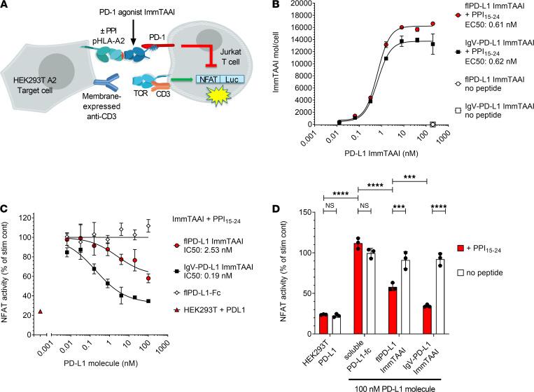
Figure 1. Target cell–bound PD-L1 ImmTAAI molecules inhibit TCR complex signaling.
(A) Schematic of the HEK293T-A2–Jurkat NFL PD-1 reporter assay. (B) PD-L1 ImmTAAI titrations were incubated with PPI15–24 peptide–pulsed or nonpulsed HEK293T-A2 target cells. ImmTAAI binding was quantified by flow cytometry, and dose-response curves were plotted (n = 3 and representative of 3 independent experiments). (C) Dose responses of the PD-L1 ImmTAAIs and PD-L1 Fc were tested in the HEK293T-A2–Jurkat NFL PD-1 reporter assay. Normalized NFAT activity was plotted against ImmTAAI concentration to calculate IC50 values (n = 3 and representative of 3 independent experiments). (D) Relative NFAT activity at 100 nM ImmTAAI was plotted for pulsed (+ PPI15–24) versus nonpulsed (no peptide) target cells (n = 3 and representative of 3 independent experiments). All data are plotted as mean ± SD and were compared by 2-way ANOVA with repeated measures and Tukey’s or Sidak’s multiple-comparison test. ***P ≤ 0.001, ****P ≤ 0.0001. IgV, immunoglobulin-like variable domain; fl, full-length.

