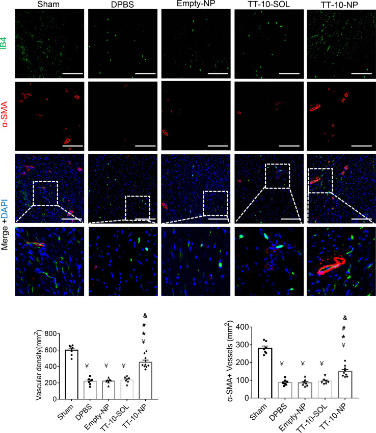
Figure 6. Assessment of TT-10–NP–mediated angiogenesis in the border zone of the infarct.
Sections from the border zone of animals that underwent MI induction, or from the corresponding regions of hearts from sham-operated animals, were obtained at week 4 and stained with the endothelial marker isolectin B4 (IB4) and for the expression of α smooth-muscle actin (α-SMA). Nuclei were identified via DAPI staining, and then vascular density and arteriole density were quantified by determining the number of IB4-positive and α-SMA–positive vascular structures, respectively, per square millimeter. Scale bar: 50 μm. n = 7–9 animals per group, 4 sections per heart, and 5 high-power fields per section. ¥P < 0.01 vs. sham, *P < 0.01 vs. DPBS, #P < 0.01 vs. Empty-NP, &P < 0.01 vs. TT-10–SOL; 1-way ANOVA with Tukey’s multiple comparisons test.

