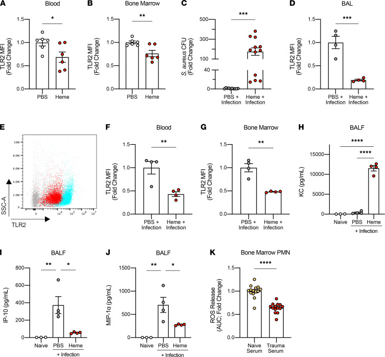
Figure 5. Heme decreases bacterial clearance in the lung and decreases TLR2 expression in PMN in vivo.
(A and B) Fold change of TLR2 MFI in PMN in the blood and the BM, 4 hours after heme challenge alone (50 mg/kg, i.p.) in vivo (n = 6/group). (C) Fold change of S. aureus CFU in BALF 24 hours after infection with and without heme challenge. Mice were challenged with heme 4 hours before the infection (50 mg/kg, i.p.; n = 10–12/group). (D) Fold change of TLR2 MFI in PMN in BAL 24 hours after infection with and without heme challenge (n = 4/group). (E) A representative flow cytometry analysis image showing the MFI of TLR2 in PMN in the BAL. Blue represents PMN in BAL from control mice, and red represents PMN from mice with heme challenge. Gray represents unstained cells. (F and G) Fold change of TLR2 MFI in PMN in the blood and the BM 24 hours after infection, with and without heme challenge (n = 4/group). (H–J) Levels of KC, IP-10, and MIP-1α in the BALF at the baseline (n = 3), 24 hours after lung infection with and without heme challenge (n = 4/group). (K) Fold change of reactive oxygen species (ROS) release in PMN treated with naive or trauma serum in response to S. aureus exposure in vitro (3 independent experiments, each with n = 3–4/group). ROS release is measured by the AUC. All data are presented as mean ± SEM. Statistical analyses for H, I, and J were performed by 1-way ANOVA with post hoc Tukey’s test. All other P values were calculated by unpaired 2-tailed Student’s t test. *P < 0.05, **P < 0.01, ***P < 0.001, ****P < 0.0001.

