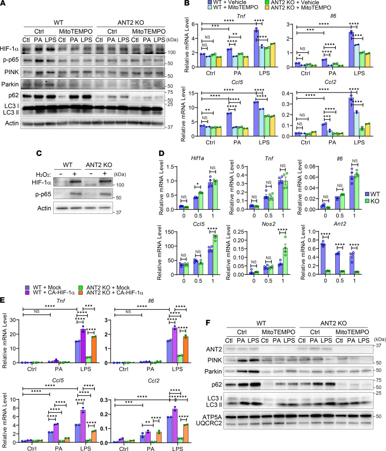
Figure 8. Increased ROS and subsequent HIF-1α stabilization mediates ANT2-dependent macrophage proinflammatory activation.
(A) Western blot analysis of inflammatory and mitophagic pathways in WT and ANT2-MKO BMDMs treated with or without PA or LPS in the presence or absence of MitoTEMPO for 6 hours. (B) mRNA expression of inflammatory genes in WT and ANT2-MKO BMDMs treated with or without PA or LPS in the presence or absence of MitoTEMPO for 6 hours (n = 2 wells/group). (C) Western blot analysis of HIF-1α and phosphorylated p65 NF-κB levels in WT and ANT2-MKO BMDMs treated with or without H2O2 for 30 minutes. (D) mRNA expression of inflammatory genes in WT and ANT2-MKO BMDMs treated with or without H2O2 for 6 hours (n = 4 wells/group). (E) mRNA expression of inflammatory genes in WT and ANT2-MKO BMDMs transfected with mock or CA-HIF-1α–expressing plasmid vector. Forty-eight hours after transfection, cells were treated with or without PA or LPS for 8 hours (n = 2 wells/group). (F) Western blot analysis of mitophagic proteins in mitochondrial extracts purified from WT and ANT2-MKO BMDMs treated with or without PA or LPS in the presence or absence of MitoTEMPO for 4 hours. In all panels, all values are mean ± SEM. *P < 0.05; **P < 0.01; ***P < 0.001; ****P < 0.0001. NS, not significant. Statistical analysis was performed by 2-way ANOVA with Tukey’s multiple comparison test.

