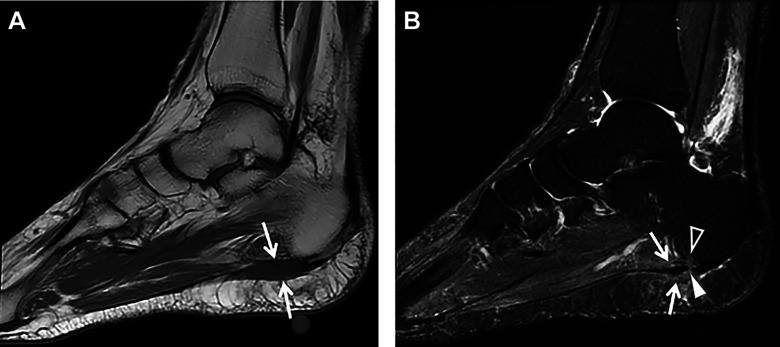
Figure 7.
(A) Sagittal T1-weighted MRI showing intermediate signal intensity in the region of the origin of the central band of the plantar fascia (arrows). (B) Sagittal STIR MRI showing heterogeneous increased signal intensity involving the proximal central limb of the plantar fascia adjacent to the vitamin E marker (arrows) consistent with plantar fasciitis. A full-thickness partial-width tear of the proximal central band of the plantar fascia is seen (arrowhead) as well as associated perifascial subcutaneous and deep soft tissue edema and a small focus of mild focal bone marrow edema in the adjacent calcaneal tuberosity (open arrowhead). MRI, magnetic resonance imaging; STIR, short tau inversion recovery.

