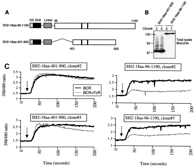
FIG. 2.
Minimal phosphatase region of SHIP alone is not sufficient for inhibition of calcium flux by FcγRIIB1. (A) Schematic diagram of the constructs designed for targeting the 401 to 900 and 96 to 1190 regions of SHIP to the FcR through fusion with the SHIP-SH2 domain via a flexible 18-amino-acid linker. (B) Expression of the constructs depicted in panel A in SHIP−/− DT40 stable clones. Equal numbers of cells were lysed, and total lysates were analyzed for expression of HA-tagged proteins by immunoblotting. Molecular size markers are indicated on the left (in kilodaltons). (C) Independent clones of SHIP−/− DT40 cells stably transfected with SH2-18aa-401-900 (left panels) or SH2-18aa-96-1190 (right panels) were loaded with indo-1 and analyzed for calcium flux as described in Materials and Methods. Recording of the fluorescence ratio was initiated prior to stimulation of cells with BCR cross-linking or BCR plus FcR co-cross-linking. The arrow indicates the time of addition of the stimulating antibody.

