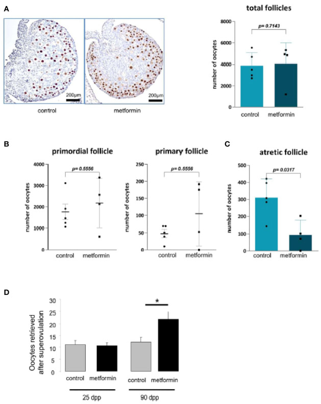
Figure 7.
Morphology of ovary at birth. (A) Number of oocytes was measured on ovary from 2 dpp old female mice after a P63 immunostaining (n=5). (B) The quantification of follicle number was performed after a staining of ovarian section with haematoxylin–eosin. Follicles were classified according to the shape and number of layers of somatic cells surrounding the oocyte: primordial (flattened cells), and primary (one layer of cuboidal cells). (C) Quantification of atretic follicles (D) and number of oocytes retrieved after superovulation at 24 dpp and 90 dpp. Scale bar= 200 µm. White arrow indicates positive cell. *p < 0.05.

