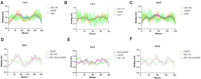
Figure 2.
Comparison between the distribution of HIV-1 IN alone or in the presence of LEDGF/p75. Either recombinant purified LEDGF/p75 or HIV-1 IN (4 nM) was incubated with chromosomes spreads under the conditions described in materials and methods section alone or together. The protein binding to chromatin was monitored by immunofluorescence as performed in Figure 1. The distribution profile of IN and LEDGF/p75 along chromosome 1, 2 and 3 was analyzed and reported respectively in (A), (B) and (C). The distribution profile of IN incubated with chromosome spreads in the presence of LEDGF/p75 has been monitored and quantified and the distribution curve has been reported in (D) for the chromosome 1, (E) for chromosome 2 and (F) for the chromosome 3. The data are reported as means from 7 to 11 serials of quantifications ± SD. In (D)–(F), the SD were omitted to improve readability.

