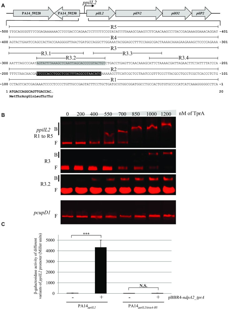
Figure 3.
TprA binds the ppilL2 promoter. (A) Promoter region of the pilL2 gene locus in the PA14 strain. The ppilL2 promoter predicted by Carter and colleagues (12) is indicated by a black box. The gray-boxed sequence from − 254 to − 287 indicates the TprA binding site (tprA-BS). The pilL2 coding sequence is in bold. (B) EMSA was performed with purified TprA protein, at concentrations ranging from 0 to 1200 nM, and different regions of ppilL2 or pcupD1 promoter (negative control). Retarded nucleoprotein complexes were identified by B (Bound); free DNA is indicated by F (Free). (C) Expression of the chromosomal ppilL2-lacZ and ppilL2ΔtprA-BS-lacZ fusions monitored in PA14ppilL2+ pBBR1MC4 or PA14ppilL2 +pBBR4-ndpa2_tprA. The strains grew in LB and the expression of two fusions were evaluated when cells reached the early stationary phase (8 h). Data are expressed in Miller units and correspond to the mean values (with error bars) obtained from three independent experiments. Wilcoxon–Mann–Whitney tests were performed and N.S. and *** indicate non-significant and P < 0.001, respectively.

