. 2021 Oct 11;12(42):14260–14269. doi: 10.1039/d1sc04671c

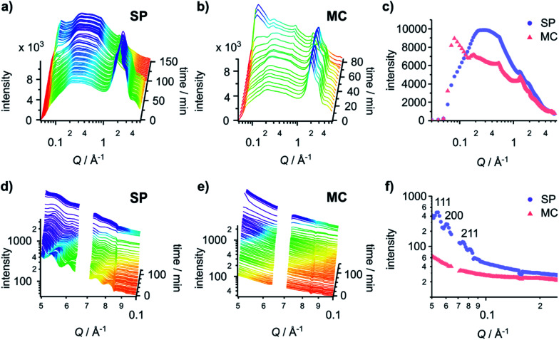
Fig. 4. Gel fibres adopt structures with different packing orders on different surfaces. (Top) time-resolved GID patterns of hydrogels formed on (a) the SP surface and (b) the MC surface. (c) The final GID patterns of the hydrogel before it fully dries out on the SP surface (103 min) and the MC surface (76 min). (Bottom) time-resolved GISAXS patterns of the hydrogels formed on (d) the SP surface and (e) the MC surface. (f) The first GISAXS patterns (at 2 min) of the hydrogel on the SP surface and the MC surface. Reflection assignments are indicated in the graph. In (d)–(f), data between Q = 0.064 Å−1 and 0.071 Å−1 was omitted as it represents a gap in the detector.

Fig. 4