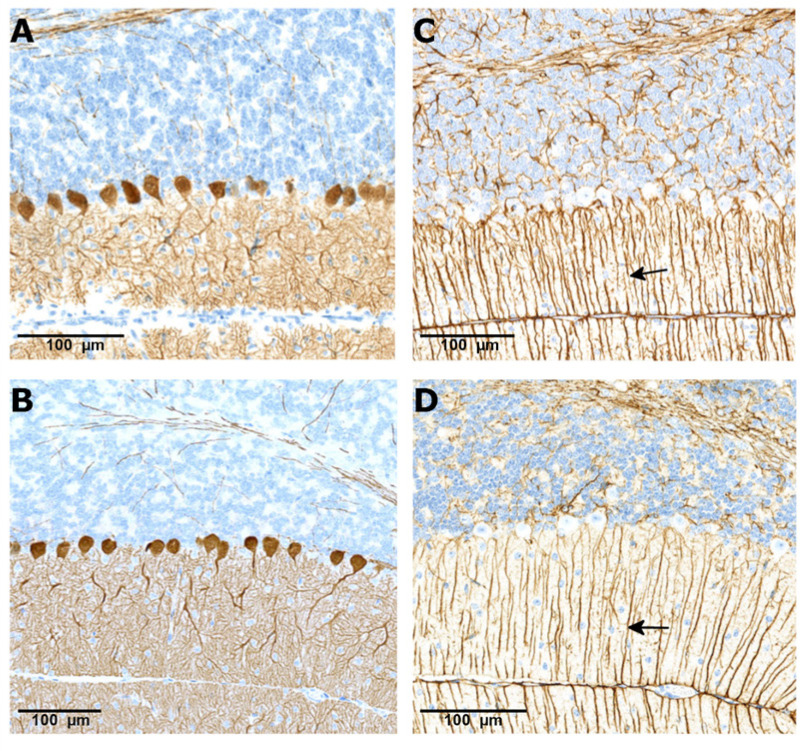
Fig. 3.
CalbindinD-28k and GFAP staining in the cerebellum of the SMA mouse model treated with vehicle and branaplam. CalbindinD-28k staining showing Purkinje cell alignment (brown staining) in a vehicle (A) treated animal at PND 14 and branaplam (B) treated animal at PND 49. Glial fibrillary acidic protein staining showing normal arrangement of Bergman glial fibers (arrows) in the vehicle- (C) and branaplam-treated animal (D). PND, postnatal day.

