Fig. 6.
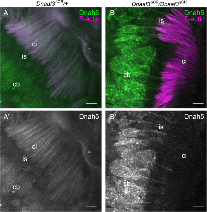
Loss of dynein heavy chain in Dnaaf3 mutant motile cilia. Immunofluorescence imaging of chordotonal neurons in pupal antenna; Dnah5 protein (green) and phalloidin staining of F-actin (red), which marks the scolopale structures surrounding the ciliary dendrites. (A) Dnaaf3ΔCR heterozygote showing Dnah5 protein in cell bodies (cb) and cilia (ci). (B) Dnaaf3ΔCR homozygote showing Dnah5 in cell bodies and dendrite inner segments (is) but absent from cilia. (A′,B′) Corresponding Dnah5 channels only. Number of antennae imaged: heterozygote n=15; homozygote n=10. Scale bars: 5 µm.

