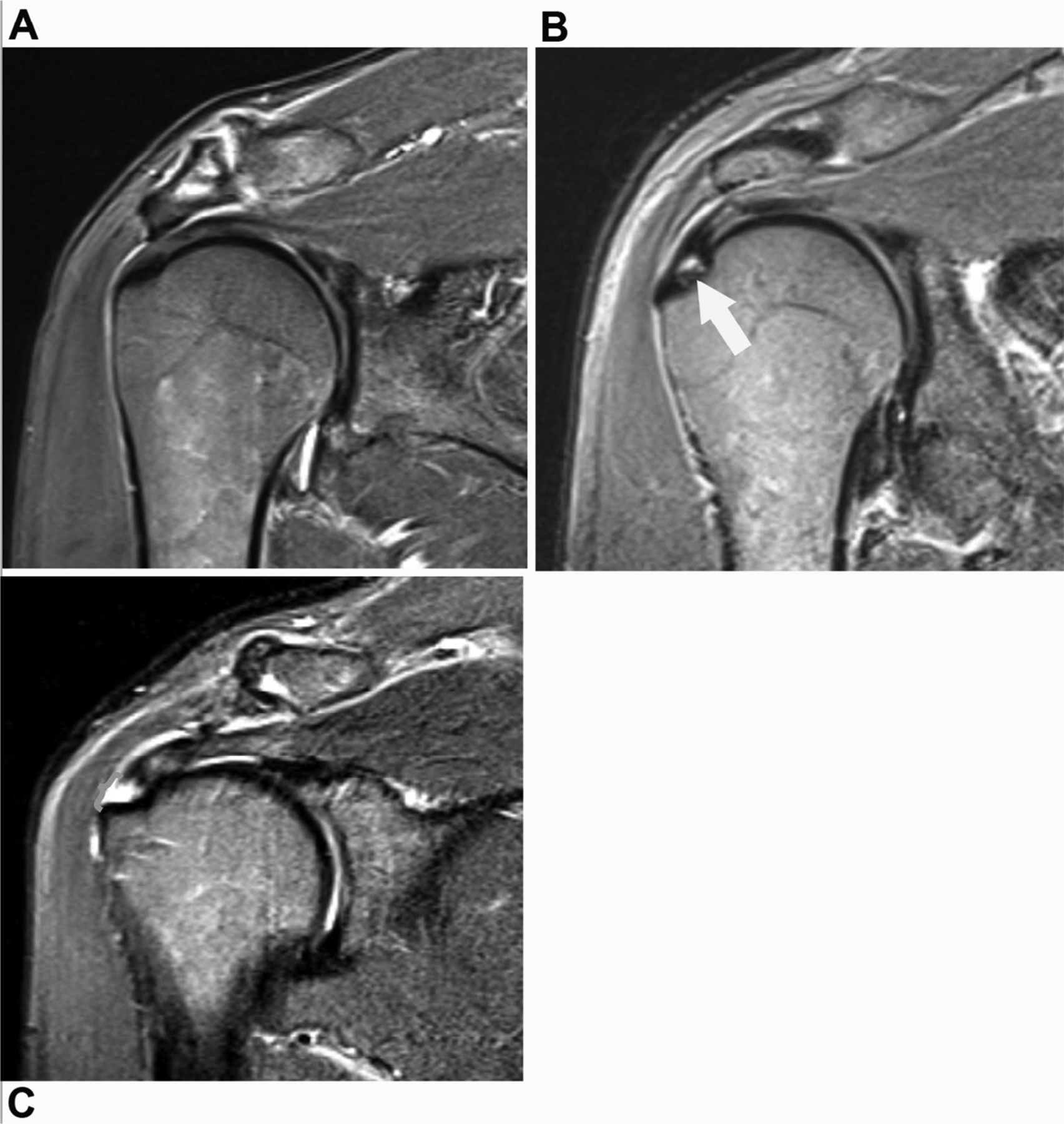
FIG 1.

Example shoulder MR images of study subjects included in each group. (A) Oblique coronal short tau inversion recovery (STIR) MR image of a 66-year-old man with no supraspinatus (SST) tear, representative of study subjects in Groups 1 and 2. (B) Oblique coronal STIR MR image of a 43-year-old man showing an partial-thickness articular-sided SST tear (white arrow), also representative of study subjects in Groups 1 and 2. (C) Oblique coronal STIR MR image of a 66-year-old man showing a full-thickness SST tear (orange bracket), representative of study subjects in Group 3. (Color version of figure is available online.)
