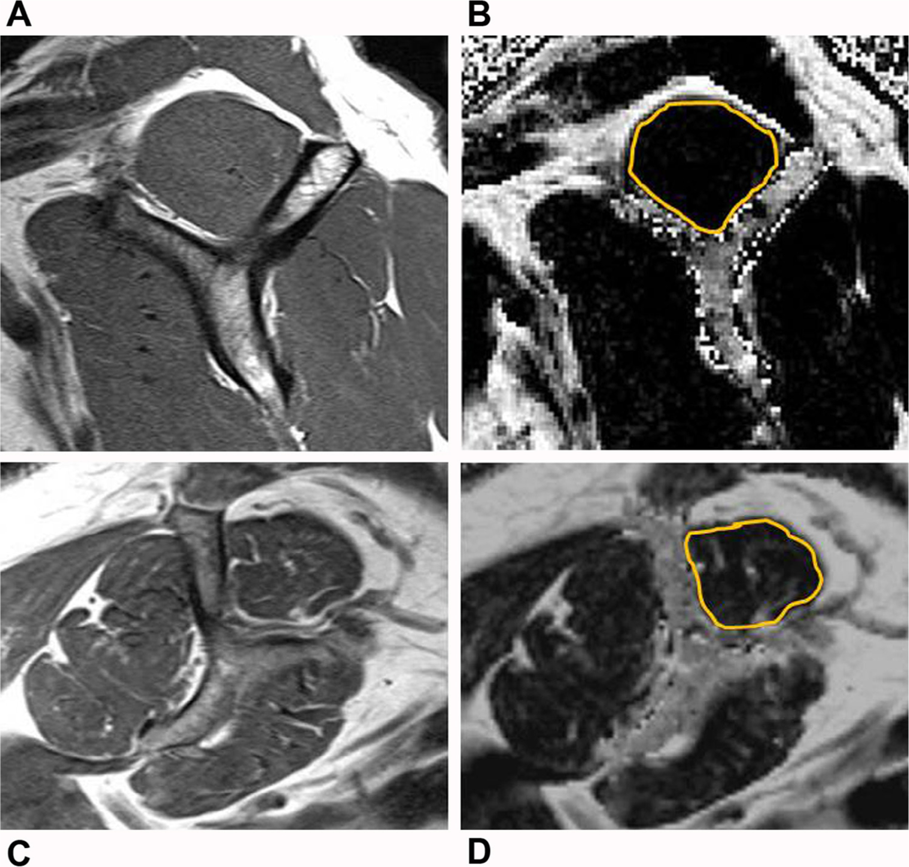
FIG 2.

Example shoulder MR images showing methods of supraspinatus muscle analysis. (A) Oblique sagittal T1-weighted MR Y-shaped view image of a 58-year-old male study subject used to determine Goutallier grade. (B) Oblique sagittal Dixon fat fraction map image, corresponding to the T1-weighted MR Y view. The gold outline denotes the manually delineated region of interest placed about the supraspinatus muscle for Dixon fat fraction analysis. (C and D) Oblique sagittal T1-weighted MR Y-shaped view and corresponding oblique sagittal Dixon fat fraction map images for supraspinatus muscle analysis in a 66-year-old female study subject.
