
Figure 2: Pretreatment setup and Identification of soft tissues using the eXIA™ 160 contrast agent.
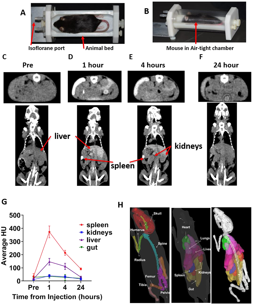
A) Custom-designed mouse holder base used in TMI treatment. B) Mouse placed in mouse holder chamber (base and transparent air-tight cylindrical chamber) to maintain continuous and homogeneous flow of isoflurane during TMI treatment delivery. C-F) The contrast agent eXIA™ 160 was injected via tail vein, and in vivo time-lapse CT imaging was carried out at different time points. Representative CT images taken at different time points are shown: C) prior to injection, D) 1h, E) 4h F) 24h after injection. G) The change in Hounsfield Units over the course of 24h after injection is shown (n=5). H) Mouse contour (3D) showing skeletal tissue and vital organs, used in developing the TMI treatment plan.