
Figure 4: Common beam arrangements and dosimetry of TMI treatment.
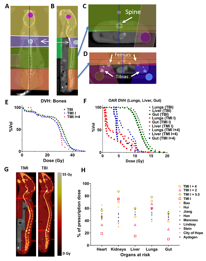
Beam arrangement for parallel opposed beams in coronal view (A), sagittal view demonstrating spine coverage (B), axial view at spine with gut avoidance (C) and axial view at pelvis with leg beams (D). A representative DVH comparing TMI and TBI plan for major organs viz., (E), bones (F) gut, liver and lung. G) Visualized dose distribution of TMI and TBI plan. H) Preclinical and clinical dosimetric comparisons: Dosimetric comparison between TMI I, TMI I+2, TMI I+4, and TMI I+5.5 (n=5), and 10 clinical TMI cases. Median dose difference represented as a percentage of the prescription dose. Regions of interest are the heart, lungs, liver, spleen, gut, kidneys, and target skeleton.