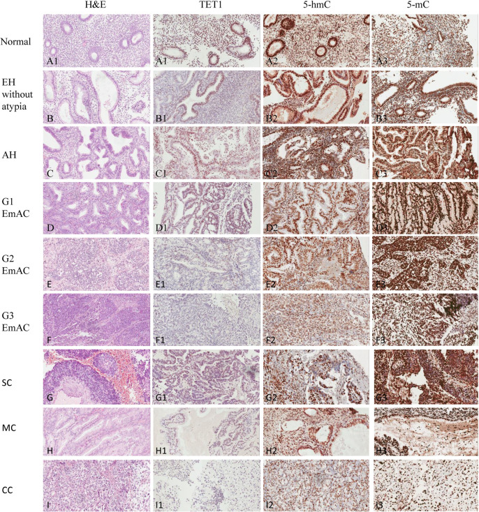
Fig 1.
H&E staining in samples of normal endometrium (A), premalignant endometrial lesions, including EH without atypia (B) and AH (C), as well as malignant samples of EC, including G1 of EmAC (D), G2 of EmAC (E), G3 of EmAC (F), SC (G), MC (H), and CC (I); TET1 expression in samples of normal endometrium (A1), EH without atypia (B1), AH (C1), G1 of EmAC (D1), G2 of EmAC (E1), G3 of EmAC (F1), SC (G1), MC (H1), and CC (I1); 5-hmC expression in samples of normal endometrium (A2), EH without atypia (B2), AH (C2), G1 of EmAC (D2), G2 of EmAC (E2), G3 of EmAC (F2), SC (G2), MC (H2), and CC (I2); 5-mC expression in samples of normal endometrium (A3), EH without atypia (B3), AH (C3), G1 of EmAC (D3), G2 of EmAC (E3), G3 of EmAC (F3), SC (G3), MC (H3), and CC (I3).

