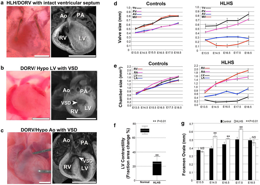
Fig. 3.
HLHS and other CHD phenotypes in the Ohia mutant line. a–c Ohia mutants with isolated hypoplasia of the aorta or LV. Some Ohia mutants exhibit DORV with hypoplasia of both the aorta and LV (a), while others can exhibit DORV with hypoplastic LV, but normal-sized Ao (b), or DORV with hypoplastic aorta, but normal LV (c). These independent occurrences of hypoplasia of the LV vs. Ao would suggest these anatomical defects are not hemodynamically driven in Ohia mutants. Scale bar, 1 mm. d–g Delineating cardiac development in Ohia HLHS mutants using fetal echocardiography. d, e Developmental profile of the growth of cardiac valves and chambers in normal vs. HLHS embryos (n = 15 embryos per group). Data shown are mean ± s.e.m. LA left atrium, LV left ventricle, RA right atrium, RV right ventricle, MV mitral valve, TV tricuspid valve, AV aortic valve, PV pulmonary valve. f Fetal ultrasound measurements show LV contractility is decreased in HLHS mutant fetuses (n = 15 embryos per group). Data shown are median with interquartile range. Wilcoxon rank-sum test, P = 0.000003. g Measurements of the foramen ovale (FO) showed FO size was not restrictive in HLHS mutants. Data shown are mean ± s.e.m. Unpaired Student’s t test: E13.5, n = 12 (control = 6, HLHS = 6) P = 0.385, t = −0.908, df = 10; E14.5, n = 18 (control = 12, HLHS = 6), P = 0.000084, t = 5.222, df = 16; E16.5, n = 27 (control = 15, HLHS = 12), P = 0.001, t = 3.7, df = 25; E17.5, n = 21 (control = 12, HLHS = 9), P = 3.4633E – 9, t = 10.262, df = 19; E18.5, n = 12 (control = 6, HLHS = 6), P = 0.453, t = −0.782, df = 10. NS not significant. Adapted with permission from Liu et al. [34]

