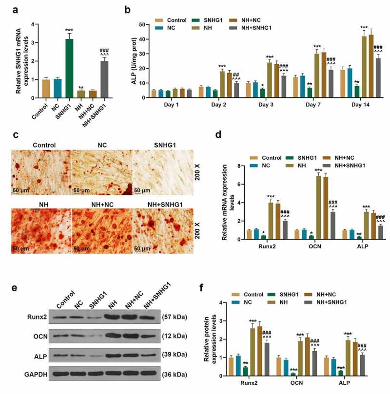
Figure 7.

SNHG1 overexpression inhibited BMSCs osteogenic differentiation. (a). The expression of SNHG1 in BMSCs after being transfected with SNHG1 overexpression plasmids or treated with NH was detected by q-PCR. GAPDH was used as an internal control. (b). The level of ALP of BMSCs after being overexpressed with SNHG1 or treated with NH was detected through microplate method. (c). The formation of calcium nodules of BMSCs after being overexpressed with SNHG1 or treated with NH was detected by Alizarin red staining. (d). The expressions of Runx2, OCN and ALP in BMSCs after being overexpressed with SNHG1 or treated with NH were detected by q-PCR. GAPDH was used as an internal control. (e-f). The expressions of Runx2, OCN and ALP in BMSCs after being overexpressed with SNHG1 or treated with NH were detected by Western blot. GAPDH was used as an internal control. All experiments were conducted in triplicate. (*P < 0.05, **P < 0.01, ***P < 0.001, vs. NC; ^P < 0.05, ^^P < 0.01, ^^^P < 0.001, vs. NH+NC; #P < 0.05, ##P < 0.01, ###P < 0.001, vs. SNHG1). (BMSCs: bone marrow mesenchymal stem cells, NC: negative control)
