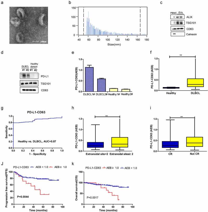
Figure 3.

Diagnostic and prognostic value of PD-L1-CD63 signal measured by SiMoa in patients with DLBCL. (a-c). Characterization of isolated exosomes by transmission electron microscopy (TEM) (a), size distribution (b) and western blots (c). (d-e) Qualification of PD-L1+ EVs in DLBCL patients and healthy subjects by western blot and SiMoa. (f) The PD-L1-CD63 signal in DLBCL and healthy subjects measured by SiMoa. (g) ROC analysis to evaluate the diagnostic power to differentiate DLBCL cases (n = 164) from the healthy controls with PD-L1-CD63 assay. (h, i) The association of PD-L1-CD63 signal with the number of extranodal sites, tumor mass and CR status after treatment. (j, k) The progression-free survival (PFS) and overall survival (OS) in high and low PD-L1-CD63 signal group
