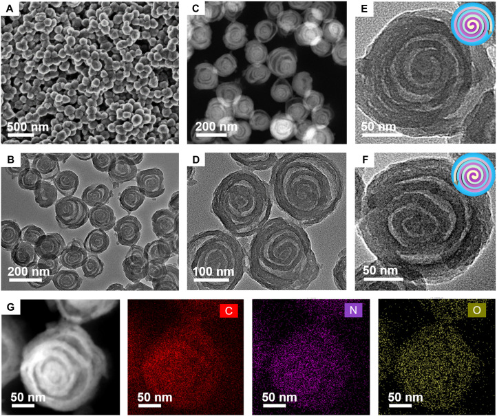
Fig. 2. Microstructure characterization of the spiral MCNs.
(A) FESEM image, (B and D) TEM image, (E and F) magnified TEM images, and (C and G) scanning TEM and energy-dispersive x-ray element mapping images of the mesoporous MCNs with unique chiral architecture prepared by the lamellar micelle spiral self-assembly strategy.

