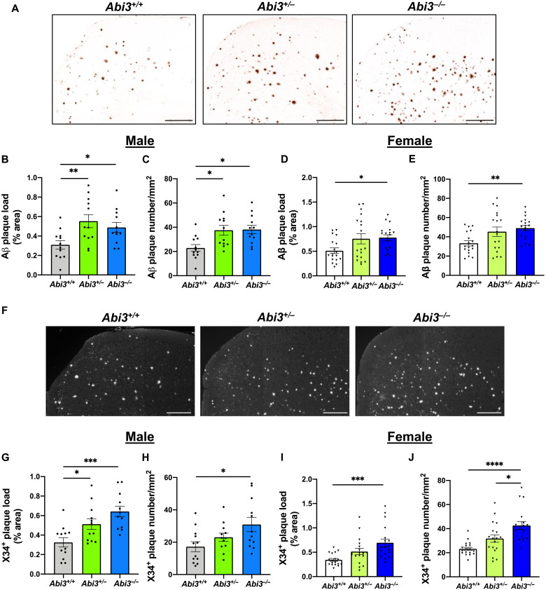
Fig. 2. Deletion of Abi3 gene locus increases amyloid plaques in 5XFAD mouse cortices.
(A) Representative images showing coronal brain sections from 8-month-old Abi3+/+, Abi3+/−, and Abi3−/− mice stained with Aβ-specific 82E1 antibody. Scale bars, 300 μm. (B) Quantification of 82E1-positive Aβ plaque area and (C) the number of Aβ plaques in the cortices of male mice (n = 12 mice per genotype). (D) Quantification of 82E1-positive Aβ plaque area and (E) the number of Aβ plaques in the cortices of female mice (n = 18 mice per genotype). (F) Representative images of brain sections from 8-month-old Abi3+/+, Abi3+/−, and Abi3−/− mice stained with X34 dye that detects fibrillar plaques. Scale bars, 200 μm. The images are converted to grayscale, and white dots represent X34+ plaques. (G) Quantification of X34+ fibrillar plaque area and (H) the number of the plaques in male mouse cortices (n = 12 mice per genotype). (I) Quantification of X34+ fibrillar plaque area and (J) the number of the plaques in female mouse cortices (n = 18 mice per genotype). Data represent means ± SEM. One-way ANOVA and Tukey’s multiple comparisons test; *P < 0.05, **P < 0.01, ***P < 0.001, and ****P < 0.0001.

