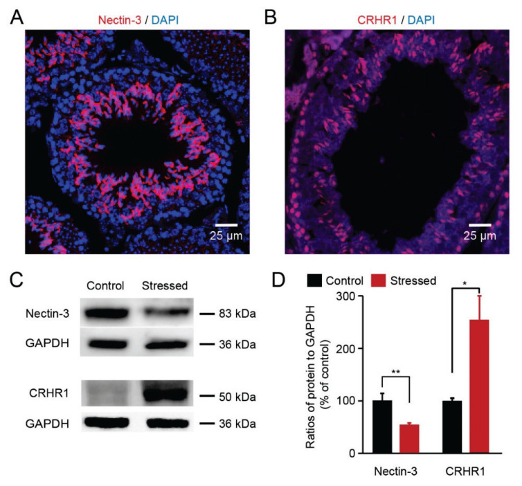
Fig. 3.
Chronic stress reduced nectin-3 protein expression and elevated CRHR1 expression (A) Immunofluorescence staining showed that nectin-3 staining colocalized with DAPI on the testis sections of each group. (B) Immunofluorescence staining showed that CRHR1 staining colocalized with DAPI on the testis sections. (C) Visualization of the protein bands of nectin-3 and CRHR1. (D) Quantitative analysis showed that nectin-3 protein levels were decreased and CRHR1 expression levels were increased in the testes of stressed mice (n=4 mice for each group, ** p <0.01, * p <0.05).

