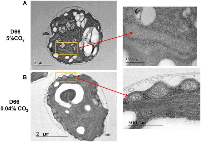
Figure 8.
Mitochondrial localization in WT D66 cells with change in CO2 levels. TEM of sectioned Chlamydomonas WT cells at (A) high CO2 and (B) ambient CO2 levels. WT cells were grown in MIN media for 48 h in high CO2 before incubating them for 12 h at their respective conditions. Areas shown by the rectangles are enlarged (right) to reveal mitochondrial structures. Scale bar, 2 µm (A), 2 µm (B), and 500 nm (enlargements).

