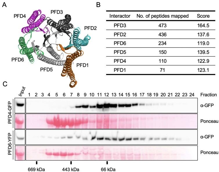
Figure 1.
The Arabidopsis PFDc. A, Predicted structure of the Arabidopsis PFDc using the human PFDc as template for modeling. B, Identification of the Arabidopsis PFDc in vivo. The table summarizes the average number of peptides and the Mascot score corresponding to each PFD subunit after TAP of GS-PFD3 (two replicates). C, Gel filtration fractions were analyzed by western blot and the fusion proteins revealed with anti-GFP antibodies. The Ponceau staining shows the large subunit of rubisco.

