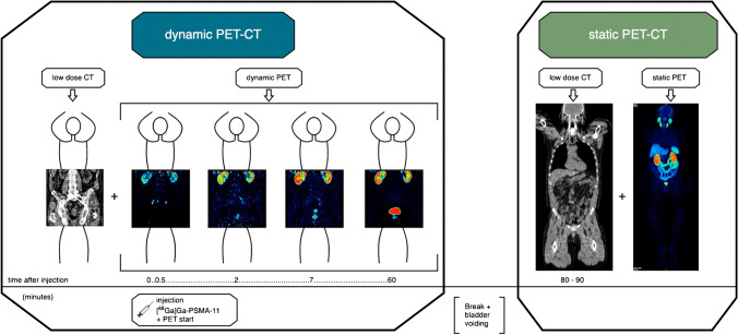
Fig. 1.
Acquisition protocol of the study. Left box: dynamic PET-CT of the pelvis and lower abdomen. After an initial low-dose CT (left image), the dynamic PET starts with injection of [68 Ga]Ga-PSMA-11 (timeline 0 min). The four PET images are arranged chronologically (left to right): after 0.5 min, 2 min, 7 min and 60 min p.i. After 60 min, the acquisition is finished and the patient goes to the toilet to empty the bladder, which is shown here as the period between the two boxes in the middle. Right box: static whole-body PET-CT 80–90 min p.i.. A low-dose CT is performed over the entire body, a part of which is shown here as an example in the left image of the right box. This is followed directly by the whole-body PET-CT image, which is shown as MIP in the right image of the right box. All images shown are in coronal slicing

