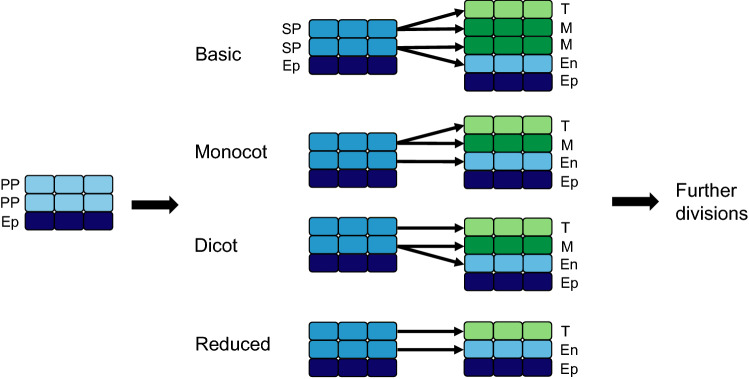
Fig. 3.
Anther wall formation types (adapted from Davis 1966). In all formation types, the epidermis (dark blue) surrounds the primary parietal cells that differentiate to form secondary parietal cells. The SP cells then differentiate into the endothecium (light blue), middle layer (dark green) and tapetum (light green), according to the formation type associated with each species. Ep: epidermis, En: endothecium, M: middle layer, PP: primary sporogenous cells, SP: secondary sporogenous cells, T: tapetum

