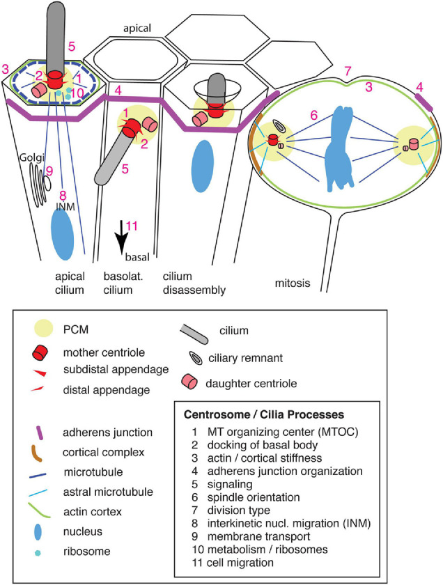
FIGURE 1.

Primary cilia and centrosomes in the ventricular zone of the developing neocortex, their structure and components. Apical progenitors exhibit an apical primary cilium, which is connected by microtubules that originate from the basal body [microtubule organizing center (MTOC)], to the cortical actin and microtubule network and the adherens junction belt. The nucleus, connected by microtubules to the centrosome, undergoes interkinetic nuclear migration (INM). Vesicles are transported along microtubules from the Golgi complex toward the apical plasma membrane and primary cilium. Newborn basal progenitors exhibit a primary cilium on the basolateral plasma membrane, which initially is still integrated in the adherens junction belt prior to delamination (arrow). Primary cilia are disassembled prior to mitosis by resorption in a ciliary pocket. During mitosis the centrosomes act as spindle poles which are anchored via astral microtubules to the cell cortex (enriched for the NuMA/LGN/Gαi complex). A ciliary remnant is localized in the vicinity of the older mother centriole. Numbers indicate the sites of the corresponding processes. Only the apical domain of the cells is depicted.
