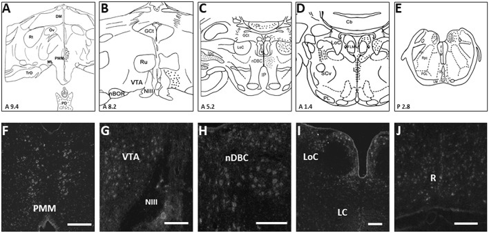
Figure 1.
Schematic drawings of rostral-to-caudal (A–E) coronal sections, showing the distribution of turkey Xenopus-like melanopsin (tOpn4x) expression labeled neurons (filled circles) of the turkey hen. Coronal illustrations were drawn from an unpublished turkey brain atlas with nomenclature taken from a chicken atlas (Kuenzel and Masson, 1988) and the revised nomenclature for avian brains (Reiner et al., 2004). Representative photomicrographs (F–J) showing the distribution of tOpn4x mRNA labeled neurons in the PMM, VTA, nDBC, LoC, LC, and R (refer to the abbreviations given below). A specific tOpn4x cRNA probe was used for in situ hybridization histochemistry (ISH). Darkfield photomicrographs of turkey brain sections processed for ISH with 33P-labeled tOpn4 antisense cRNA probes. Scale bar: 100 μm (G,H,J), 200 μm (F,I). The following abbreviations are used in the figure: Cb, cerebellum; DM, dorsomedial hypothalamic nucleus; MLF, medial longitudinal fasciculus; GCt, mesencephalic central gray; Ipc, parvocellular nucleus Isthmi; Imc, magnocellular nucleus isthmi; IP, interpeduncular nucleus; LC, caudal linear nucleus; LM, medial lemniscus; LoC, locus coeruleus; ML, lateral mammillary nucleus; nBOR, nucleus of the basal optic root; nDBC, nucleus decussationis brachiorum conjunctivorum; NIII, third cranial nerve; nTS, nucleus of the solitary tract; Ov, nucleus ovoidalis; PD, pars distalis; PH, plexus of Horsley; PL, lateral pontine nuclei; PMM, premammillary nucleus; R, raphe nucleus; Rpc, parvocellular reticular nucleus; Rt, nucleus rotundus; Ru, nucleus ruber; SCv, nucleus subcoeruleus ventralis; TrO, tractus opticus; VTA, ventral tegmental area (Modified from Kang et al., 2010).

