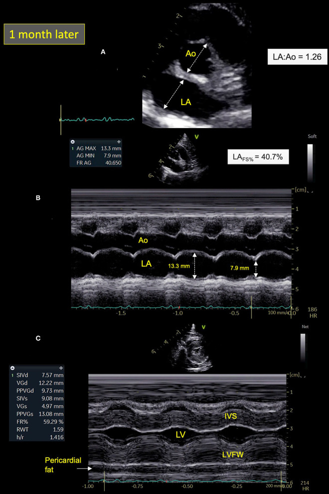
Figure 4.
Two-dimensional (2D) right parasternal short-axis view (A) and M-mode echocardiograms (B,C) from Cat-1 1 month after initial presentation. (A) As compared with day 0 (see Figure 2A), this 2D right parasternal short-axis view taken at end-diastole at the level of the aortic valve shows a markedly decreased left atrial size (left atrium:aorta ratio (LA:Ao) = 1.26 vs. 2.09) associated with disappearance of spontaneous echo contrast, pleural effusion, and pulmonary consolidation. (B) This M-mode image obtained from the 2D right parasternal short-axis view taken at the level of the aortic valve confirms normalization of the LA fractional shortening (LAFS% = 40.7 vs. 8.4%). (C) This M-mode echocardiogram obtained from the right parasternal transventricular short-axis view confirms marked asymmetric left ventricular hypertrophy, more pronounced for the left ventricular free wall (LVFW) than the interventricular septum (IVS), i.e., end-diastolic thicknesses of 9.7 and 7.6 mm, respectively [normal values <6 mm (22)]. Note also posteriorly the important amount of pericardial fat (maximal thickness of 6 mm). LV, left ventricle.

